Eye in Orbit, Magnified 5 Times, in 7 Parts
Product Code : SCL-PL-12340
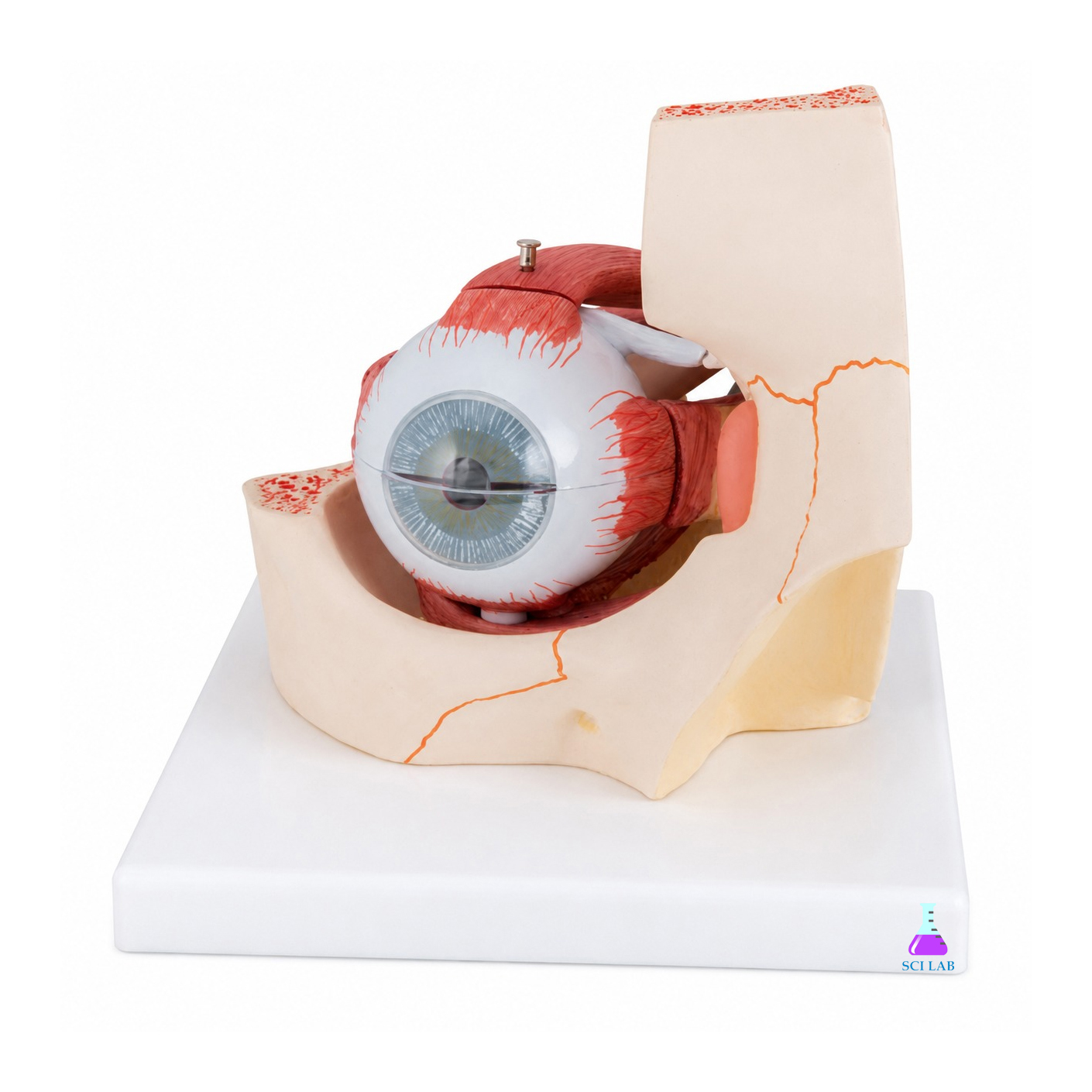
Eye in Orbit, Magnified 5 Times, in 7 Parts (SKU SCL-PL-12340) is an enlarged model of the human eyeball set in the bony orbit, which opens up into its layers so students can see the cornea, iris, lens, retina and vitreous body. It serves Class 10 and 12 biology, optometry, nursing and medical anatomy teaching. The model is a Science Lab Supplies product, manufactured at the Saha, Ambala plant in India and quoted straight to schools, colleges, tender contractors and distributors.
Specifications
Scale, parts, base, weight and size of the 5x eye-in-orbit model each take a row below. In short, the eye is shown at five times natural size and divides into seven parts: the upper sclera with cornea and muscle insertions, two choroid halves with iris and retina, the lens and the vitreous body, all on an orbit-bone base.
| Specification | Value |
|---|---|
| SKU | SCL-PL-12340 |
| Magnification | 5 times natural size |
| Parts | 7 |
| Sclera | Upper half removable, with cornea and eye muscle insertions |
| Choroid | Both halves removable, with iris and retina |
| Lens | Crystalline lens, removable |
| Vitreous body | Removable |
| Base | Orbit bone |
| Weight | 1.24 kg |
| Dimensions | 18 x 18 x 20 cm |
| Manufacturer | Science Lab Supplies, Saha, Ambala, Haryana |
| Country of origin | India |
Applications and Experiments for the Eye in Orbit Model
Taking the eye apart layer by layer shows how light passes through the cornea and lens to the retina, and where the eye muscles attach. The orbit base places the eyeball in its bony socket, which helps when teaching the eye as part of the skull rather than as an isolated organ.
- Class 10 science: the human eye, its parts and how it forms an image, compared with the convex and concave lenses of the Glass Lens Demonstration set.
- Class 12 physics and biology: the eye as an optical instrument and its defects, which the Human Eye kit Model (Functional) can demonstrate with its flexible lens.
- Optometry and ophthalmic assistant courses: layers of the eyeball and extraocular muscles.
- Nursing and MBBS anatomy: the orbit and the structure of the eye.
Standard Scope of Supply
The model is supplied as one assembled unit on its orbit-bone base, with all seven parts shown in the table fitted in place, ready to be taken apart in class. Any optional items you add, such as charts or other sense-organ models, appear as separate lines on the quotation.
Packing, MOQ and Lead Time
You can ask us to quote one eye model for a teacher's desk or a number for several biology and optometry classrooms. Our quotation shows the packing, delivery time and price for exactly that quantity, and the charts or other anatomy models below can be included.
Science Lab Supplies holds an Importer-Exporter Code as a registered exporter, so it can ship to buyers in Egypt, Iraq, Nigeria, Kenya, Ghana and Ethiopia. The business is registered under Udyam as a manufacturing enterprise (UDYAM-HR-01-0014546) and for GST (GSTIN 06ADEPJ0574F2ZU). The number of eye models per classroom can be sent through the contact page or the enquiry cart; sense-organ models in a bid go via the lab tender page.
Frequently Asked Questions
Which parts of the eye model can be removed?
The upper sclera half, both choroid halves, the crystalline lens and the vitreous body lift out, making seven parts with the orbit base, so each layer of the eye can be shown and returned in order.
How big is the model?
It is five times natural size, measures 18 x 18 x 20 cm and weighs 1.24 kg, so it can be seen clearly from the front rows of a classroom.
Who makes the Eye in Orbit model?
This eye model is manufactured by Science Lab Supplies at its own factory in Saha, Ambala, India. Buyers deal with the maker directly, and the factory itself prepares each quotation without a trading intermediary.
Is the model suitable for teaching eye defects?
It shows the cornea, lens and retina in position, which gives teachers a clear base for explaining how focusing errors arise, alongside ray diagrams drawn on the board.
Related Eye, Head and Anatomy Models
Other eye models show the organ at a different scale or with its eyelids, while skull, ear, skeleton and torso models and a laminated chart place it in the wider body. All of these items can be priced on one quotation with the eye model for a biology or optometry classroom.
- Human Eye Model, Enlarged 3 Times — six parts on a stand, numbered with a key card.
- Human Eye Model with Eyelids — horizontally sectioned eye with removable sclera and muscles.
- Human Ear Model, 3 Parts — 4–5 times enlarged ear with removable tympanic membrane.
- Human Skull Model — shows the teeth of the upper and lower jaw.
- Teaching Boards — laminated 50 x 67 cm charts, including a human eye sheet.
- Stan Skeleton, on Metal Stand with 5 Castors — skull assembled in 3 parts with inserted teeth.
- African Torso, Bisexual, in 24 Parts — torso with the head in 3 parts.
Browse the full Physics Lab range, the Head and Facial Model range or all physics lab equipment.
Last updated: 22 October 2026


.png)