
Animal Cell Model
Product Code : LSK-BL-10219
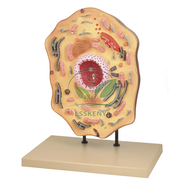
Animal Cell Model (SKU LSK-BL-10219) is a detailed model of an animal cell enlarged 20,000 times, showing the nucleus, endoplasmic reticulum, mitochondria, Golgi apparatus, centrioles, ribosomes, lysosomes and fat vacuoles. It suits cell-biology lessons from middle school to Class 11 and first-year college biology, where students can name each organelle on one large form. Science Lab Supplies quotes the model from Ambala, India, for schools, colleges, tender contractors and distributors building a set of biology models for schools.
Specifications
The table sets out each specification of this 20,000-times enlargement. In short, the model shows eight animal cell structures, from the nucleus and endoplasmic reticulum to the centrioles and fat vacuoles, weighs about 2,350 g and measures approximately 380 x 240 x 460 mm, so every organelle can be pointed out during a lesson.
| Specification | Value |
|---|---|
| SKU | LSK-BL-10219 |
| Subject | Animal cell |
| Magnification | Enlarged 20,000 times |
| Nuclear and synthesis structures | Nucleus, endoplasmic reticulum, ribosomes, Golgi apparatus |
| Other organelles shown | Mitochondria, centrioles, lysosomes, fat vacuoles |
| Weight | Approx. 2,350 g (2.35 kg) |
| Dimensions | Approx. 380 x 240 x 460 mm |
| Supplied by | Science Lab Supplies, Ambala, India |
Applications and Experiments for the Animal Cell Model
The model can be used to teach the parts of an animal cell and the job of each organelle, from protein assembly on ribosomes to energy release in mitochondria. It suits NCERT Class 8 and Class 9 cell chapters, Class 11 cell biology and B.Sc. first-year cytology, making it useful school biology lab equipment in India.
- Class 8–9 science: identifying the nucleus, mitochondria and vacuoles before students look at real cells on an Animal Tissue Prepared Microscope Slide.
- Class 11 biology (Cell: The Unit of Life): relating the endoplasmic reticulum, ribosomes, Golgi apparatus and lysosomes to protein synthesis, packaging and digestion.
- Plant and animal cell comparison: centrioles appear on this model, while plant cell models add a cell wall and plastids.
- Cell division lessons: locating the centrioles before showing chromosome stages on the Cell Division slide set of 9 or the Meiosis Model 3D Cell.
Standard Scope of Supply
Each order line covers one Animal Cell Model enlarged 20,000 times, showing the eight structures named in the table, at about 2,350 g and 380 x 240 x 460 mm. The model is supplied as a single unit as described above; any optional items are listed on the quotation.
Packing, MOQ and Lead Time
Quotations can be requested for a single model for one classroom or for several models across a school group or college department. Each quotation from Science Lab Supplies gives the packing, delivery time and price for that quantity, and slides, charts or other cell models can be added to the same document.
Holding an Importer-Exporter Code, Science Lab Supplies is able to ship to buyers in Bangladesh, Zambia, Rwanda, South Africa, Qatar and Sri Lanka. For a price on this cell model, send a message through the contact page, place it in the enquiry cart beside your other biology items, or use the lab tender page for institutional bids.
Frequently Asked Questions
What does the Animal Cell Model show?
It shows the nucleus, endoplasmic reticulum, mitochondria, Golgi apparatus, centrioles, ribosomes, lysosomes and fat vacuoles of an animal cell, enlarged 20,000 times so that each structure can be named and compared during a lesson.
How large and heavy is the model?
It measures approximately 380 x 240 x 460 mm and weighs about 2,350 g, or roughly 2.35 kg. Check the shelf or display space you have before ordering several models for one room.
How does it differ from the plant cell models in the range?
This model carries animal cell features such as centrioles and fat vacuoles, while the Plant Cell Model is a 20 cm diameter model of plant cell structures. Ordering both lets students compare the two cell types side by side.
Who supplies the Animal Cell Model?
Science Lab Supplies, based in Ambala, Haryana, India, quotes and dispatches this model as a supplied item for schools, colleges, training institutes and distributors, with the packing and delivery time stated on each quotation it sends.
Related Cell Models and Charts
These models and teaching aids cover the same unit on cell structure: plant cell forms, a protozoan cell, cell organelles, cell division and a wall chart. Any of them can be listed on one quotation with this model for a complete cell-biology display.
- Plant Cell Model — 20 cm diameter model for exploring plant cell structures.
- Plant Cell on Base Model — electron-microscope view with part of the wall removed to show vacuoles, plastids and mitochondria.
- Model of the Plant Cell — colour-coded PVC model, 180 x 300 x 60 mm, on a base with numbered key card.
- Superstructure of the Cell (With Cell Organelles) — PVC model of cell organelles measuring 700 x 400 x 500 mm.
- Amoeba Cell Structure Model — magnified Amoeba proteus cell with contractile and food vacuoles and pseudopodia.
- Enlarged 3D Mitosis Model — multi-stage model of the phases of somatic cell division.
- The Structure of the Animal Cell — laminated wall chart, 70 x 100 cm.
Browse the full Biology Lab range, the General Biology Products range for protozoan and cell-division models, or all biology lab equipment.
Last updated: 9 October 2026


