
Eye Ball Model
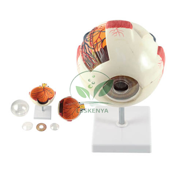
Product Code : LSK-BL-10151
Eye Ball Model (SKU LSK-BL-10151) is an eyeball enlarged about three times that separates in a horizontal plane into 6 parts, revealing the ocular muscles, blood supply, cornea, iris, ciliary body, retina, optic disc, vitreous body and sclera. It sits on a 150 mm pedestal stand and comes with a key card for anatomy and sense-organ lessons. Science Lab Supplies quotes the model from Ambala, India, for institutions, resellers and tender buyers.
Specifications
The table records each feature of the pedestal eyeball separately. In short, it is roughly 3x natural size, splits horizontally into six parts, displays nine named structures from the ocular muscles and cornea to the retina, optic disc and vitreous body, stands on a 150 mm high pedestal and includes a key card.
| Specification | Value |
|---|---|
| SKU | LSK-BL-10151 |
| Scale | Enlarged about 3 times |
| Number of parts | 6 |
| Plane of separation | Horizontal |
| Shows | Ocular muscles |
| Shows | Blood supply |
| Shows | Cornea and sclera |
| Shows | Iris and ciliary body |
| Shows | Retina and optic disc |
| Shows | Vitreous body |
| Mounting | Pedestal stand, 150 mm high |
| Key card | Supplied |
| Supplied by | Science Lab Supplies, Ambala, India |
Applications and Experiments for the Eye Ball Model
The eyeball can be used to show the layers of the eye and the path of rays to the retina: once the horizontal halves are lifted apart, students can trace the cornea, iris, ciliary body, vitreous body and retina in order. It suits secondary science, senior biology and nursing anatomy classes.
- Class 10 science (The Human Eye and the Colourful World): the role of the cornea, iris and ciliary body in focusing, and correcting short sight with a Concave Optics Experiment Lens.
- Class 11 biology (Neural Control and Coordination): the retina, optic disc and blind spot.
- Following the visual pathway to the occipital lobe shown on the Human Brain Model- 2 Parts.
- Nursing and paramedical anatomy: the ocular muscles and blood supply of the eyeball.
Standard Scope of Supply
The Eye Ball Model is supplied with its 150 mm pedestal stand and a key card naming the structures on the six parts. Lenses for optics demonstrations, wall charts and other eye models are not included, and any of them can be quoted on the same document if required.
Packing, MOQ and Lead Time
Whether the order is for one pedestal eyeball or a set for every biology group, Science Lab Supplies issues a quotation that names the packing, delivery time and price for that quantity. Lenses or other sense-organ models can be quoted in the same reply for convenience.
Science Lab Supplies, an Importer-Exporter Code holder, can ship to buyers in the Philippines, Nepal, Bangladesh, Zambia, Rwanda and South Africa. Requests for this eyeball can be sent from the contact page or gathered in the enquiry cart, and buyers working to a tender can refer to the lab tender page.
Frequently Asked Questions
Which structures does the Eye Ball Model show?
It shows the ocular muscles, blood supply, vitreous body, optic disc, ciliary body, iris, cornea, retina and sclera. The model separates horizontally into 6 parts so the inner structures can be seen.
How is the model mounted?
The eyeball sits on a pedestal stand 150 mm high, which keeps it raised for viewing from the front of the class. A key card identifying the structures comes with it.
How does it compare with the Human Eye Model?
Both are about three times life size and have six parts. The Eye Ball Model uses a 150 mm pedestal and key card, while the Human Eye Model has a detachable base, a photocopiable key and a 90 x 90 x 150 mm size.
Who supplies the Eye Ball Model?
Enquiries for the Eye Ball Model are handled by Science Lab Supplies in Ambala, India. It prepares quotations and arranges dispatch for schools, colleges, distributors and tender contractors, including mixed orders of eye and ear models.
Related Human Eye Models
Each of these eye models adds something different, such as eyelids and orbit, a functional lens or a larger scale, so they work well next to the pedestal eyeball when comparing anatomy and vision. Any combination can be priced together on one quotation.
- Human Eye Model — dissectable into 6 parts, 90 x 90 x 150 mm, with photocopiable key.
- Functional Eye Model, Non-Liquid — retina screen slides along the optical axis to simulate myopia and hyperopia.
- Human Eye Model, Enlarged 3 Times — both halves of sclera with cornea and eye muscle attachments separate.
- Human Eye Model with Eyelids — horizontally sectioned eye in the orbit with intraocular muscles.
- Eye in Bony Orbit Model — two of the six eye muscles are removable.
- Eye in Orbit, Magnified 5 Times, in 7 Parts — crystalline lens and vitreous body removable, on the orbit bone.
- Human Eye kit Model (Functional) — flexible lens focuses an image on the retina; 17 x 17 x 22 cm.
Explore the Biology Lab range, the Head and Facial Model range, or all biology lab equipment.
Last updated: 9 October 2026


.jpg)
